

Summary: NIST researchers have developed a novel infrared imaging method that enables the observation and quantification of biomolecules in living cells, overcoming previous limitations caused by water absorption.
Estimated reading time: 7 minutes
An innovative infrared imaging technique developed by researchers at the National Institute of Standards and Technology (NIST) has opened up new possibilities for observing biomolecules inside living cells. This method, known as solvent absorption-compensated infrared microscopy (SAC-IR), overcomes a long-standing challenge in cellular imaging by eliminating interference caused by water absorption.
The research team, led by NIST chemist Young Jong Lee, has successfully used this technique to measure the absolute mass of proteins and other biomolecules in individual cells over time. This breakthrough could accelerate progress in several fields, including drug development, cell therapy and biomanufacturing.
Why it matters: This new imaging method provides a noninvasive way to study cellular processes in real time, without the need for potentially harmful dyes or markers. By offering a more precise and standardized approach to measuring biomolecules in cells, SAC-IR microscopy could lead to significant improvements in our understanding of cell biology and the development of new medical treatments.
How to overcome the problem of water in infrared images
Infrared microscopy has long been a powerful tool for identifying and analyzing chemical structures in various materials. However, its application in living cells has been limited due to the strong absorption of infrared light by water, which makes up a large portion of cellular contents.
Dr. Lee explains the challenge: “In the spectrum, water absorbs infrared rays very strongly, and we want to see the absorption spectrum of proteins through the thick water background, so we designed the optical system to uncover the contribution of water and reveal the signals of proteins.”
The SAC-IR technique uses a specialized optical element to compensate for water absorption, effectively eliminating the “noise” created by water and allowing researchers to focus on signals from other biomolecules. This is similar to using a sun-blocking filter to view an airplane flying close to the sun: by removing the overwhelming light source, the object of interest becomes visible.
Observation of cellular processes in real time
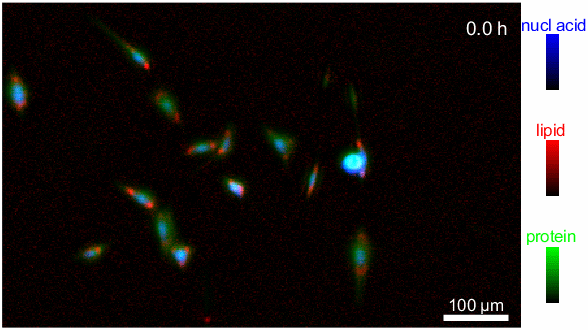
Using a custom-designed IR laser microscope equipped with the SAC technique, the NIST team was able to observe fibroblasts (cells that support the formation of connective tissue) over a 12-hour period. This extended observation allowed the researchers to identify and measure various biomolecules, including proteins, lipids, and nucleic acids, during different stages of the cell cycle.
Although 12 hours may seem like a long time, it is actually a significant improvement over current alternatives. Many existing techniques require the use of large synchrotron facilities, which are not easily accessible to most researchers and have limited time to conduct experiments.
Implications for biomedical research and industry
The ability to measure absolute amounts of biomolecules in individual living cells has far-reaching implications for both basic research and applied biotechnology. Some potential applications include:
- Cancer cell therapy: SAC-IR could help assess the safety and efficacy of modified immune cells used in cancer treatments. “In cancer cell therapy, for example, when you modify cells from a patient’s immune system to better recognize and destroy cancer cells before reintroducing them into the patient, you have to ask yourself, ‘Are these cells safe and effective? ’ Our method may be useful by providing additional information regarding biomolecular changes in cells to assess cellular health,” Lee said.
- Drug development: The technique could speed up drug discovery and testing by allowing researchers to measure how different cell types react to potential drug candidates at the molecular level.
- Cell preservation: By providing detailed information on cellular composition, SAC-IR could help optimize cell freezing and thawing processes, which is crucial for long-term cell storage in medical and research applications.
- Standardization of cellular measurements: The ability to quantify biomolecules consistently across different laboratories could lead to more reproducible results in cell biology research.
Future directions and challenges
While the current implementation of SAC-IR microscopy represents a significant advance, the NIST team is already looking to expand its capabilities. Future research aims to improve the accuracy of measurements for specific biomolecules such as DNA and RNA.
Researchers also hope to use this technique to answer fundamental questions in cell biology, such as identifying biomolecular signatures that correspond to different stages of cell viability. This could have important implications for fields such as regenerative medicine and tissue engineering.
As with any new technology, scaling up the SAC-IR technique is likely to present challenges to its widespread use. Issues such as cost, ease of use, and integration with existing laboratory workflows will need to be addressed. However, the potential benefits of this new imaging method make it an exciting development in the field of cell biology and biomedical research.
Proof:
- What is the main challenge that SAC-IR microscopy overcomes in cellular imaging?
- How long were the researchers able to observe the fibroblast cells using the new technique?
- Name a possible application of SAC-IR microscopy in biomedical research.
Answer key:
- The strong absorption of infrared light by water in cells, which previously obscured other biomolecules.
- 12 hours
- Possible answers include: evaluating modified immune cells for cancer therapy, drug development and testing, optimizing cell preservation methods, or standardizing cell measurements in laboratories.