


Skeletal muscle contraction is fundamental to all types of movements in animals, from simple locomotion to complex manipulations. This contraction is driven by contractile proteins within muscle cells, particularly actin and myosin, which are organized into myofibrils. Myofibrils are further divided into sarcomeres, the functional units of muscle contraction. While actin forms the thin filaments, myosin constitutes the thick filaments, playing a pivotal role in the sliding filament model of muscle contraction. Myosin is not limited to muscle tissue; it also contributes to several cellular functions in non-muscle cells, such as cell adhesion and migration.
In this article, we will explore the structure, synthesis, classification, and various functions of myosin, focusing on its crucial roles in muscle contraction and intracellular processes.
Structure of myosin
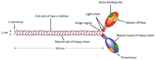
Myosin is a filamentous protein classified as a motor protein due to its ability to convert chemical energy from ATP hydrolysis into mechanical work. A single myosin molecule is composed of six subunits: two heavy chains and four light chains. The structural organization of these chains is essential for its function.
- Heavy chains:The two heavy chains coil around each other to form a double helix, which forms the tail of the myosin molecule. This tail forms the bulk of myosin's structure.
- Myosin headAt one end of the heavy chains, they separate and form globular structures known as myosin heads or cross-bridges. Each head contains an ATPase site and actin binding sites.
- Light chains:The globular heads are associated with two light chains each, which stabilize the structure of the heads.
In general, the myosin molecule consists of two heads and a tail, forming a distinctive structure necessary for its function.
Myosin domains
To understand the function of myosin, it is helpful to consider its three main domains:
- Main domain:This domain is globular and is formed by the end of the heavy chain and two light chains. It is responsible for binding to actin filaments and has ATPase activity crucial for muscle contraction.
- Neck dominanceThe neck domain, which acts as a link between the head and tail, is essential for transducing the force generated by the heads to the tail. It also binds light chains.
- Tail controlFormed by the coiled-coil structure of heavy chains, the tail domain connects myosin molecules within a filament and interacts with cargo molecules in non-muscle cells.
Myosin synthesis
Myosin synthesis is a complex process involving multiple steps of gene expression.
Transcription
The process begins with transcription, in which the DNA sequence of a myosin gene is copied into messenger RNA (mRNA). This occurs in the nucleus of muscle and non-muscle cells. Each gene corresponds to a specific myosin isoform, and only one gene is transcribed at a time.
Post-transcriptional modifications
To prepare mRNA for translation, several modifications occur:
- 5' CapA guanosine triphosphate (GTP) cap is added to the 5' end of the mRNA to protect it from degradation and aid in the initiation of translation.
- Poly-A glueA polyadenylated tail is added to the 3' end, which further protects the mRNA and aids in its export to the cytoplasm.
Translation
Once in the cytoplasm, the mRNA is translated into a myosin protein. Ribosomes assemble around the mRNA, and transfer RNA (tRNA) molecules carry amino acids to the ribosome according to the mRNA sequence. This process continues until a stop codon is reached, which signals the end of translation. The newly synthesized myosin protein then undergoes post-translational modifications in the endoplasmic reticulum.
Post-translational modifications
Post-translational modifications are essential for the functional maturation of myosin:
- Phosphorylation:The addition of phosphate groups to serine, threonine, or tyrosine residues, catalyzed by myosin light chain kinases, can activate or deactivate myosin function.
- Nitration and Nitrosylation:Addition of nitrate or nitro groups can occur under pathological conditions, affecting myosin function and potentially leading to contractile dysfunction.
Myosin classes
Myosin is classified into several types based on its structure, location, and function. The main classes include:
- Myosin I:A monomeric protein involved in intracellular transport and membrane interactions.
- Myosin II:Classical muscle myosin responsible for muscle contraction, present in skeletal, smooth and cardiac muscles.
- Myosin III:It is found in the eyes of Drosophila and is involved in light-dependent transduction.
- Myosin V:A dimeric protein that “walks” along actin filamentscrucial for intracellular transport.
- Myosin VI:Responsible for the transport of endocytic vesicles within cells.
- Myosin VII:It participates in phagocytosis and spermatogenesis and is found in some sensory structures.
- Myosin VIII:Present in plant cells, regulating cell division and cytoplasmic flow.
- Myosin XI:A dimeric protein involved in the movement of organelles within plant cells.
Role in muscle contraction
Myosin plays a central role in muscle contraction through interactions with actin filaments. The mechanism varies slightly between skeletal, smooth, and cardiac muscles.
Skeletal muscle
In skeletal muscle, myosin filaments are located at the center of sarcomeres, and actin filaments extend from each end. Myosin heads bind to actin filaments when the binding sites are exposed due to the release of calcium ions. ATP hydrolysis drives the conformational change in the myosin heads, resulting in a power stroke that pushes the actin filaments toward the center of the sarcomere. This filament-sliding mechanism causes muscle contraction.
Smooth muscle
Smooth muscle contraction is regulated differently. Myosin filaments are interspersed with actin filaments attached to dense bodies. Unlike skeletal muscle, smooth muscle lacks troponin and tropomyosin. Instead, contraction is regulated by phosphorylation of myosin light chains by myosin light chain kinase, activated by calcium ions. This process allows myosin heads to bind to actin and facilitate contraction.
Cardiac muscle
Cardiac muscle contraction follows a mechanism similar to that of skeletal muscle, with myosin filaments arranged in sarcomeres. The sliding filament model also applies here, where calcium ions trigger the contraction process.
Summary
Myosin is a protein that is essential for muscle contraction and various cellular processes. Its structure, composed of two heavy chains and four light chains, is essential for its function. Myosin synthesis involves transcription, translation, and post-translational modifications. Myosin is classified into different types based on its function and location, including muscle and non-muscle forms.
In muscle contraction, myosin interacts with actin filaments via a sliding filament mechanism. While the fundamental process is similar across muscle types, the regulatory mechanisms differ between skeletal, smooth, and cardiac muscles. Understanding the structure and function of myosin provides insight into its diverse roles in both muscle physiology and cellular processes.


Leave feedback about this