


Muscles are composed of specialized proteins that enable them to contract and produce movement. Among these proteins, actin and myosin play crucial roles in muscle contraction. They work together to facilitate voluntary and involuntary movements in humans and other animals. Actin and myosin are critical to the functioning of muscle cells and are also involved in several cellular processes. This article explores the differences between actin and myosin, highlighting their roles, structures, and functions in muscle contraction.
Actin
Definition and structure
Actin is a globular protein, known for forming thin filaments in muscle cells. In its monomeric form, it is known as G-actin (globular actin). When polymerized, G-actin forms filamentous actin (F-actin), which is a major component of the cytoskeleton in eukaryotic cells. Actin filaments are crucial for maintaining cell shape, enabling cell motility, and facilitating several cellular processes such as division and intracellular transport.
Actin filaments are about 7 nanometers in diameter and vary in length, typically between 2 and 2.6 micrometers in muscle cells. The filaments are composed of two intertwined strands of actin molecules, giving them a helical structure.
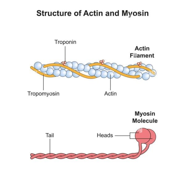
Regulatory proteins
Actin filaments are associated with several regulatory proteins, including tropomyosin and troponin. Tropomyosin binds along the actin filament groove and blocks myosin binding sites, whereas troponin, which binds to tropomyosin, regulates the interaction between actin and myosin. The binding of calcium ions to troponin causes a conformational change that moves tropomyosin away from the binding sites, allowing myosin to bind to actin and initiate contraction.
Location
In muscle cells, actin filaments are present in both the A (anisotropic) and I (isotropic) bands of the sarcomere. The I band contains only actin filaments, whereas the A band contains both actin and myosin filaments. The actin filaments are anchored to the Z-disc at the ends of the sarcomere.
Appearance and surface
Actin filaments have a smooth surface. When viewed under a microscope, they have clearer striation patterns than myosin filaments. The smoothness of the actin surface is due to the uniform arrangement of G-actin subunits along the filament.
Function
Actin filaments function primarily to maintain cell shape, facilitate movement, and interact with myosin to produce muscle contractions. During contraction, actin filaments slide toward the H zone of the sarcomere, which is the central region of the A band.
Abundance
Actin filaments are more numerous than myosin filaments in muscle cells. For each myosin filament, there are approximately six actin filaments, reflecting their relative abundance.
Myosin
Definition and structure
Myosin is a motor protein characterized by its ability to convert the chemical energy of ATP hydrolysis into mechanical energy. It forms thick filaments in muscle cells and plays a central role in muscle contraction. Myosin proteins are composed of heavy chains and light chains, with the heavy chains forming long, rod-shaped tails and globular heads that interact with actin filaments.
Myosin filaments are thicker and longer than actin filaments, measuring approximately 15 nanometers in diameter and 4 to 5 micrometers in length. The filaments are composed of multiple myosin molecules arranged in a bipolar structure, with the heads projecting outward.
Regulatory proteins
Myosin filaments are associated with meromyosin, which includes the light chains and head domains that interact with actin. The head regions of myosin contain ATPase activity, which is crucial for the conversion of ATP into mechanical energy.
Location
Myosin filaments are predominantly found in the A bands of the sarcomere. They overlap with actin filaments in the A band, but are not present in the I band.
Appearance and surface
Myosin filaments have a rough surface due to the presence of protruding myosin heads. They have a darker appearance in striation patterns compared to actin filaments, which contributes to the darker appearance of the A band.
Function
Myosin filaments do not slide into the H zone during contraction, but instead form cross-bridges with actin filaments, facilitating the sliding of actin filaments towards the center of the sarcomere. This sliding mechanism is essential for muscle contraction.
Abundance
Myosin filaments are less abundant than actin filaments. Normally, a myosin filament interacts with several actin filaments, reflecting their lower abundance in muscle cells.
Muscle contraction is facilitated by the sliding filament model, which describes how actin and myosin interact to produce movement. When a motor neuron stimulates a muscle, calcium ions are released from the sarcoplasmic reticulum. These ions bind to troponin, causing a displacement of tropomyosin that exposes actin binding sites.
Myosin heads, activated by ATP hydrolysis, bind to exposed sites on actin to form cross-bridges. The myosin heads then rotate and pull the actin filaments toward the center of the sarcomere. This action shortens the sarcomere, resulting in muscle contraction. The process is repeated as long as ATP and calcium ions are available.
Key differences between actin and myosin
| Aspect | Actin | Myosin |
| Definition and function | Globular protein that forms thin filaments; involved in muscle contraction, cell shape, motility and division. | Motor protein; converts ATP hydrolysis into mechanical energy; forms thick filaments and interacts with actin for muscle contraction. |
| Structure | Thin filaments, approximately 7 nm in diameter; helical structure made of G-actin. | Thick filaments, about 15 nm in diameter; consisting of a long rod-shaped tail and globular heads. |
| Size | Short (2-2.6 µm long), thin (0.005 µm diameter). | Long (4-5 µm in length), thick (0.01 µm in diameter). |
| Surface characteristics | Smooth surface. | Rough surface due to protruding myosin heads. |
| Regulatory proteins | Tropomyosin (blocks myosin binding sites); Troponin (binds calcium and regulates tropomyosin position). | Meromyosin (includes head and tail domains, involved in cross-bridge formation). |
| Location in the Sarcomere | It is found in the A and I bands; anchored to Z disks. | Found mainly in A bands; anchored on the M line. |
| Abundance | Most abundant; typically six actin filaments per myosin filament. | Less abundant; one myosin filament for every six actin filaments. |
| Cross-bridge formation | It does not form cross-bridges directly; it provides binding sites for myosin. | Forms cross-bridges with actin filaments during contraction. |
| Partnership with ATP | It is not directly associated with ATP. | Directly associated with ATP; ATPase activity drives movement. |
| Sliding mechanism during contraction | It slides into the H zone during contraction. | It remains stationary while pulling actin filaments toward the center of the sarcomere. |
| Ends and binding | One free end (barbed or positive end), the other end attached to the Z disk (pointed or negative end). | Both ends free; heads remain associated with ATP. |
| Appearance under microscope | It appears as lighter stretch marks (I bands). | It appears as darker stretch marks (A bands). |
| Additional Features | Forms microfilaments in the cytoskeleton; participates in cell division and motility. | It functions as a molecular motor in muscle contraction and other cellular processes depending on the type of myosin. |
Conclusion
Actin and myosin are essential for muscle function and a variety of cellular processes. Actin forms thin filaments that provide structural support and interact with myosin during contraction. Myosin, as a motor protein, forms thick filaments and is essential for generating the force required for muscle movement. Understanding the differences between these two proteins highlights their distinct roles in muscle physiology and their cooperative function in the mechanism of muscle contraction.
Leave feedback about this